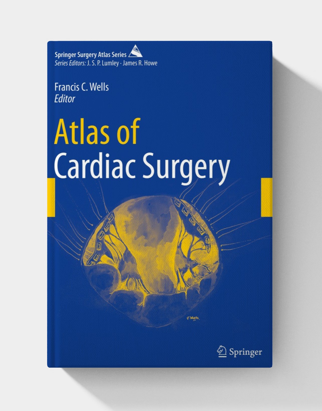
By: Francis C. Wells
PDF: 24.1
This Atlas is illustrated with rich pictures of cardiac surgical specimens. It not only contains normal heart specimens but also dissects those specimens, taking pictures from various angles to create a three-dimensional representation. It also includes reviews of the specimens’ pathological reviews. Chapters 1 through 10 introduce the normal anatomy of the cardiac chambers and surgical approaches to the heart, while chapters 11 through 28 describe 18 kinds of congenital heart defects. There are a total of over 1,000 images and illustrations in this book, which will be of great interest not only to the surgeons but also to the cardiologists, anesthesiologists, and surgical pathologists.
PDF Preview
Description
How to use ?


How to Use
Once you purchase the product, we will share a Google Drive link with you via email. Through this link, you will gain access to:
The downloadable PDF.
Any additional content, such as videos or quizzes, if included.
Simply click the link, download the materials to your device, and start exploring at your own pace.


